INCLUSIONS IN GRANULOMAS
 | ||||||
| ||||||
A variety of inclusions may be found in granulomas, usually within the cytoplasm of giant cells. They are non-specific findings which may be characteristic of one or another entity but are without diagnostic significance.
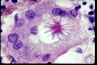
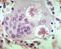
ASTEROID BODIES: Stellate inclusions with numerous rays radiating from a central core. May be seen in granulomas of various entities but are most frequently encountered in the giant cells of foreign body granulomas. Structures strongly resembling asteroid bodies may be seen rarely in the cytoplasm of tumor giant cells and in fibrin-rich exudates.
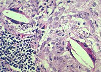
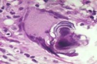
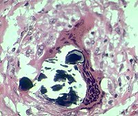
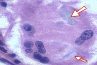
CRYSTALLINE INCLUSIONS: Colorless refractile crystals composed predominantly of calcium oxalate are frequently found in the giant cells of granulomas of sarcoidosis and other diseases. In some cases they may serve as the nidus for deposition of calcium leading to formation of Schaumann (conchoidal) bodies (Figs. 5,6)
| ||||||
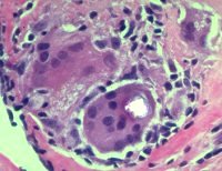
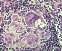
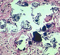
SCHAUMANN (CONCHOIDAL) BODIES: Large concentric calcifications often containing refractile calcium oxalate crystals (Figs. 5-7). Although usually intracytoplasmic they may, if numerous or very large, be extruded into the extracelular space (Fig. 11).
1. Asteroid body, sarcoidosis
2. Asteroid bodies, foreign body granuloma
4. Calcium oxalate crystal-polarized
5. Early Schaumann body forming around calcium oxalate crystals, polarized
| ||||||
| ||||||
| ||||||
9. Schaumann body
7. Fragmented Schaumann.body surrounding calcium oxalate crystals (at arrows). Polarized
8. Schaumann body
10. Large fragmented Schaumann body
11. Extracellular Schaumann bodies
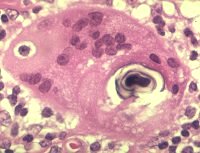
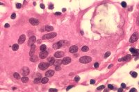
SLIT-LIKE (CHOLESTEROL) CRYSTALS: These are occasionally seen in granulomas of various types.
| ||||||
| ||||||
| ||||||
12. Cholesterol crystals
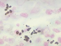
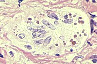
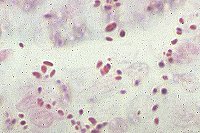
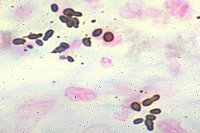
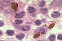
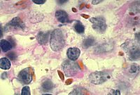
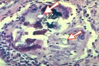
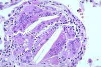
HAMAZAKI-WESENBERG (H-W) BODIES: Although these structures may occasionally occur as inclusions within giant cells (Fig. 17 ), they are usually found extracellulary in the sinusoids of granuloma-containing lymph nodes in sarcoidosis as well as in non-granulomatous lymph nodes from patients with sarcoidosis and other conditions.
They are oval or spindle-shaped, range up to several microns in size, and appear yellow-brown with H&E stain (Figs. 14-17). The pigment appears to be lipofuscin. Ultrastructural studies have shown that H-W bodies are giant lysosomes and residual bodies. They are acid-fast and stain with silver stains as well as a variety of other stains.
When visualized with silver stains H-W bodies can be easily mistaken for budding yeasts due to clustering of individual bodies (Figs. 18,19).
| ||||||
| ||||||
| ||||||
| ||||||
| ||||||
| ||||||
14. H-W bodies-lymph node-40X
15. H-W bodies-lymph node - oil immersion
16. H-W bodies-lymph node - oil immersion
17. H-W bodies in giant cell-lung
18. H-W bodies-lymph node - methenamine silver -oil immersion. Note resemblance to budding yeasts.
19. H-W bodies-lymph node - methenamine silver -oil immersion. Note resemblance to budding yeasts.
| ||||||
13. Cholesterol crystals
| ||||||
3. Calcium oxalate crystals, H&E
| ||||||
6. Early Schaumann body forming around calcium oxalate crystals.